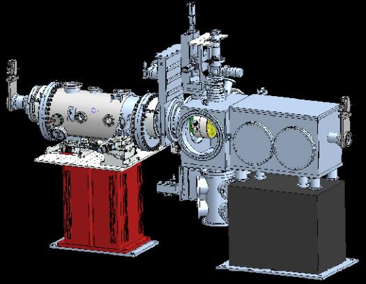
CXI Components
Some CXI components are located in the X-ray Tunnel, roughly 60 m upstream of the CXI hutch. These devices do not need to be close to the sample and were placed away in a less crowded area.
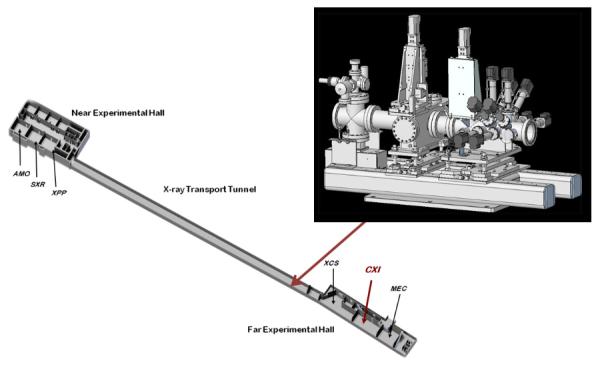
The attenuator system with 10 individually actuated filters is the first component upstream, followed by the Pulse Picker and the X-ray Focusing Lenses. Most downstream is a pumping system. The X-ray Focusing Lenses are located 60 m upstream of the CXI Sample Chamber in order to produce a 10 µm FWHM focal spot.
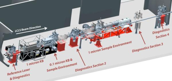
Optics
A single pulse shutter that can be used to allow only a single FEL pulse to pass through to the experimental chambers.
A millisecond shutter from is incorporated into a vacuum chamber on a translation stage to allow insertion into the beam. It can be operated up to 30 Hz.
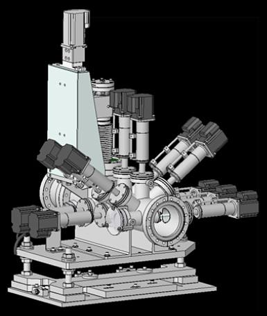
A set of silicon foils of varying thicknesses is used to tailor the intensity of the LCLS beam. Multiple attenuation factors will be possible by introducing any desired combination of foils into the LCLS beam path.
Ten foils of different from 20 microns to 10.24 mm thicknes is used to provide close to 3 steps of attenuation per decade in the 6-15 keV range.
Cylinders of 3 mm diameter made of silicon nitride (Si3N4) or Tantalum are used to slit the beam. Silicon nitride does not get damaged by the LCLS beam downstream of the Near Experimental Hall and can be used to cut into the main part of the beam.
In the case of the CXI instrument, these slits will primarily be used to remove unwanted intensity around the central FEL beam to produce a cleaner beam.
The tantalum slits have a much higher absorption than the Si3N4 and can be used to slit down the harmonics of the beam efficiently up to 25 keV.
Diagnostics
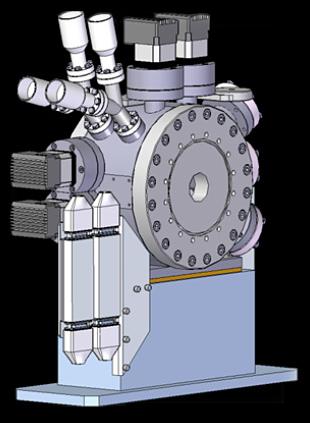
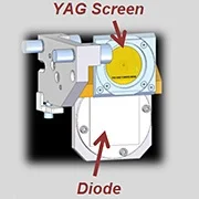
The spatial profile of the LCLS beam is measured at various locations along the CXI beamline using a scintillating screen and a high resolution camera-lens combination.
The screen is mounted on a translation stage to allow insertion into the beam.
The profile measurement is destructive of the beam, meaning the beam terminates at these devices and is used for alignment and troubleshooting procedures.
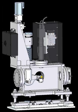
The intensity of the LCLS beam can be measured at various locations along the CXI beamline using a photodide which is mounted on a translation stage to allow insertion into the beam.
The intensity measurement is destructive of the beam, meaning the beam terminates at these devices and is used for alignment and troubleshooting procedures.
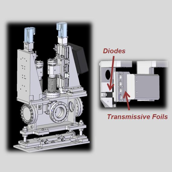
A thin foil allowing most of the beam to be transmitted is used to measure the LCLS pulse energy on a shot-to-shot basis. Compton back-scattering from the thin foil is measured using a set of diodes located upstream of the foil.
The sensing area of the diodes is facing the foil and they are placed in a tiled arrangement leaving a hole in the middle. The integrated intensity of all the diodes provides a measurement of the beam intensity on every pulse.
The relative signal from each tile can be used to get a measurement of the beam position.
The Wavefront Monitor is a scintillating screen and a high resolution camera-lens combination.
The screen is mounted on a 2-axis translation stage to allow insertion into the beam and potentially centering of a beam stop or attenuator right in front of the screen.
The beam profile can be measured downstream of the experiment and can be used to measure low angle scattering.
Algorithms can in principle be used to recover the wavefront from the measured beam profile downstream of the focus.
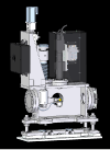
CXI Reference Laser
While the LCLS beam is unavailable to the CXI instrument either during machine studies or because the beam is being used by a different instrument, it is possible to align the CXI experiment using a laser beam collinear with the LCLS beam.
The green laser beam is introduced into the vacuum beamline using a vacuum window and an in-vacuum mirror.
Window valves are provided downstream of the Reference Laser System to allow the laser beam to be used with any part of the beamline vented to air.
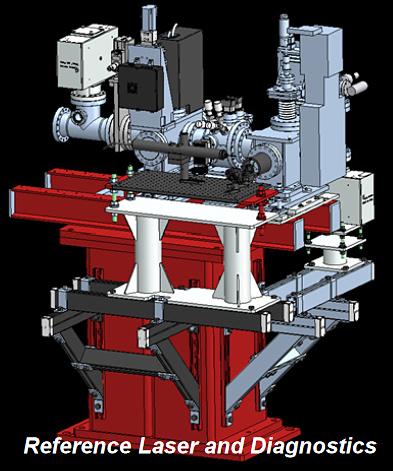
Focusing Optics
Compound refractive lenses made of Beryllium can be used to produce a 10 µm focus in the CXI 1 micron Sample Chamber. A translation stage allows one of three stacks of lenses to be selected which allows certain ranges of photon energies to be focused.
Not all photon energies canl be focused by the lenses since the are chromatic optics.
These lenses are installed in the X-ray Tunnel 57 m upstream of the CXI Sample Chamber. The lenses are only usable for photon energies above 4 keV due to the high absorption below this energy.
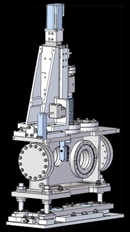
A second set of lenses can be installed inside the CXI hutch roughly 3.2 m upstream of the 1 micron Sample Chamber on Diagnostics Section 2 (DG2).
These lenses can be used with the unfocused beam to produce a ~3 x 3 micron focus at the sample.
The same lenses can also be used along with the 1 micron KB mirrors to provide a flexible spot size at the sample on the same axis as the KB beam by moving the focal plane of the KB-focused beam.
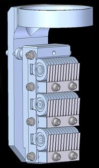
A set of lenses can be installed on the detector chamber downstream of the CSPAD detector.
Since the detector has a hole in the middle, the "spent" beam passing through it can be captured by lenses and refocused in a second sample chamber downstream.

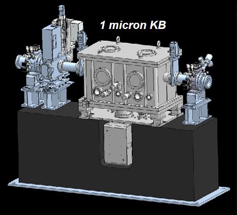
A pair of elliptical mirrors are used in a Kirpatrick-Baez configuration to produce a diffraction limited spot of roughly 1 micron at the interaction region of the CXI instrument (inside the 1 micron Sample Chamber).
The midpoint between the KB pair is located 8.5 meters upstream of the focus and this produces a round focal spot between 1 and 4 microns FWHM, depending on the photon energy.
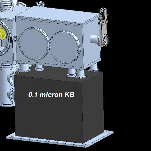
A pair of elliptical mirrors are used in a Kirpatrick-Baez configuration to produce a diffraction limited spot of roughly 0.1 micron at the interaction region of the CXI instrument (inside the 0.1 micron Sample Chamber).
The midpoint between the KB pair is located 0.7 meters upstream of the focus and this will produces in principle an elliptical focal spot of roughly 90 x 150 nm2 FWHM at 8.3 keV.
The spot size varies with photon energy since the source size varies with photon energy and the mirrors aperture the beam at lower photon energy.
The focal spot is expected to be on the order of 400 nm at 2 keV.
Diagnostics Section 2 (DG2)
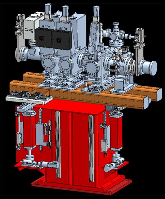
A gate valve is followed by Slits, and then X-ray Focusing Lenses. An Intensity-Position Monitor allows the beam intensity to be measured and is followed by a Profile-Intensity Monitor.
A differential pumping system is placed just before the downstream gate valve.
Detector Stage Location B (DSB)
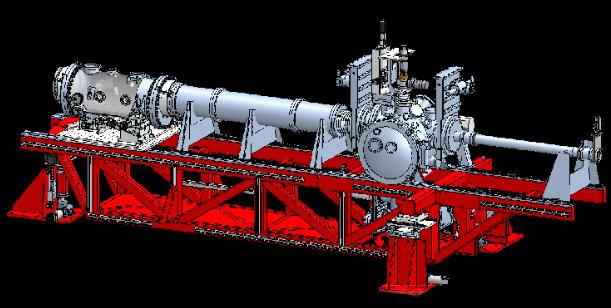
The CXI Detector Stage can be placed on the upstream side of the CXI 1 micron Sample Chamber. It can be used for backscattering measurements.
This is a rare occurence and for the majority of the time, this section of the beamline is used for multiple optics and diagnostics.
Typically, slits, attenuators, a pulse selector and a time tool are located on the DSB section.
CXI Sample Chambers
A large vacuum chamber capable of supporting various experimental configurations is used to control the environment of the sample. The pressure inside the chamber is typically on the order of 10-4 to 10-7 Torr depending on the samples .
Inside the chamber can be multiple options for aperture and sample stages, including a 5-axis sample piezo stage and different configurations of stepper motor stages. Samples mounted on thin supports can be positioned accurately into the LCLS beam using a long range microscope with on-axis viewing of the sample.
The sample environment will also allow samples to be delivered to the LCLS beam without a substrate, in the gas phase by interfacing with the CXI particle injector or any other sample delivery system that can connect to a 6 inch Conflat flange.
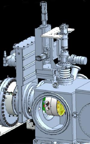
The 1 micron Sample Chamber is the main sample Chamber for CXI and can be used with the 1 µm KB focus, the beam focus with either sets of X-ray Focusing Lenses, with the unfocused beam or using the refocused spent beam from the 100nm KBs.
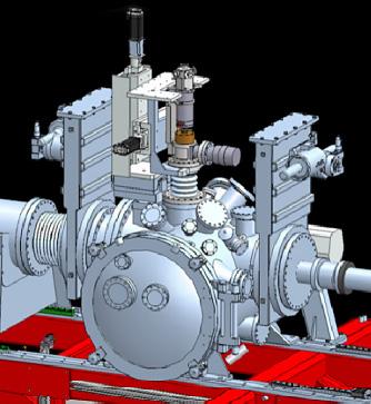
The 0.1 micron Sample Environment has all the functionality of the 1 micron Sample Environment except that this sample environment is only usable with the 100 nm KB focus.
Sample Delivery
A single particle injector will be used to transfer particles from the gas phase at atmospheric pressure into the high vacuum of either Sample Environments.
A tightly focused beam of particles will be delivered and the Particle Injector will be mounted on translation stages allowing the beam to be steered into the interaction region.
These translation stages can be used with any other sample delivery system which interfaces to a 6 inch Conflat flange.
The injector provided by CXI requires highly concentrated samples to provide a high hit rate due to the random arrival time of the particles at the interaction region.
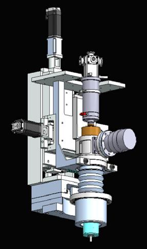
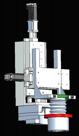
A mechnical system compatible with multiple liquid jet delivery system is available for use on both sample chambers aty CXI.
This mechanical system was originally developed at ASU.
Sample Diagnostics
A small ion time-of-flight mass spectrometer can be used to detect the creation of ions at the interaction region.
These ions produced by the explosion of the sample when hit by an LCLS pulse can be accelerated away from the interaction region and their arrival time can be detected.
This can provide a trigger signal when an injected particle was hit and also provide some information on the chemical composition of the particle that was hit.
2D Detector System
A 2D X-ray pixel array detector is used to measure the scattered x-rays, in particular in the forward scattering geometry. The detector consists of 4 tiles with a central hole which is fixed in size at 9mm.
The Jungfrau4M has 2048 x 2048 pixels. The Jungfrau detector was developed but the Paul-Scherrer Insitut.
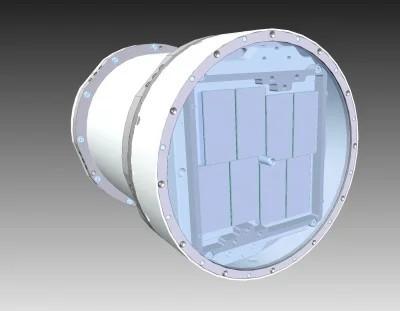
A 2D X-ray pixel array detector is used to measure the scattered x-rays. The detector is made of several tiles arranged so that a central hole is present.
The central hole size can be varied from 1.5 to 9.4 mm. Each quadrant of the detector moves as a solid unit.
The CSPAD has 1516 x 1516 pixels. Two full size CSPADs are available for use at CXI. A CSPAD is the dedicated detector for the parasitic SSC sample chamber.
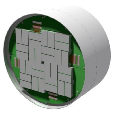
The 2D X-ray detector is mounted in vacuum and it is accurately positioned relative to the interaction region and the LCLS beam direction. It is possible to vary the sample to detector distance from 50 mm to 600 mm for the front detector.
The back detector can move from 2000 to 2600 mm. Only the front detector can be used with the 100 nm sample chamber while both front and back are available for use with the 1 micron sample chamber.
The Detector Stage positions the detector transverse to the LCLS beam so that the direct beam passes through the central hole in the detector. A 500 mm stage in-vacuum allows continuous variation of the detector distance over that range without breaking vacuum.

CXI Precision Instrument Stand
The 1 micron Sample Environment is mounted on a large frame with 6-axis positioning.
This frame is almost 6 m long and capable of supporting many tons and can be used to attach user supplied equipment.
The CXI Precision Stand provides a very flexible platform for user needs.

CXI Sample Environments
The 1 micron Sample Environment includes the 1 micron Sample Chamber, the Particle Injector and the Detector Stage which can be mounted at multiple locations.

The 0.1 micron Sample Environment has all the functionality of the 1 micron Sample Environment except that this sample environment is only usable with the 100 nm KB focus.
The detector distance is limited to 50-600 mm for this setup.
