MEC Diagnostics & Components
The following tabs provide an overview of X-ray beamline components, in-house experimental diagnostics, and standard optical beam delivery platforms at MEC. Additional technical details, CAD drawings and analysis methods (when available) for the components presented below may be found on the MEC Confluence website (under construction). An overview of the MEC instrument is published in a 2015 JSR article.
XRD diffraction
X-ray Diffraction can be performed with the use of ePix10k detectors arranged around the interaction point to reach the desired 2θ and φ coverage. MEC has 4 specialized ePix10k designated for diffraction diagnostic in a high EMP environment, having superior shielding to standard ePix10k quads, and for higher photon energies, having 2x thicker silicon sensors (1 mm). The detectors are mounted on dedicated holders allowing removal and kinematic repositioning of the detector on its mount. This also allows the detectors to be mounted substantially differently from the standard configuration arrangement.
VISAR
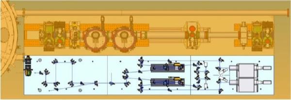
A line-imaging velocity interferometer system for any reflector (VISAR) is a widely used optical interferometric diagnostic for dynamic (e.g. shock) experiments. For opaque targets, VISAR is capable of determining shock speeds by detecting shock breakout times as a function of target thickness. In addition, free surface expansion velocities can be determined from measurement of the phase introduced in the probe beam due to the surface motion. The VISAR in the MEC station has spatial resolution of 10 µm and a temporal resolution of 10 ps, with time window ranging from 1 ns to 1 ms, a field of view of 1 mm, and a minimal velocity per fringe of 0.5 km/s/fringe.
Von Hamos X-ray Thompson Scattering Spectrometers
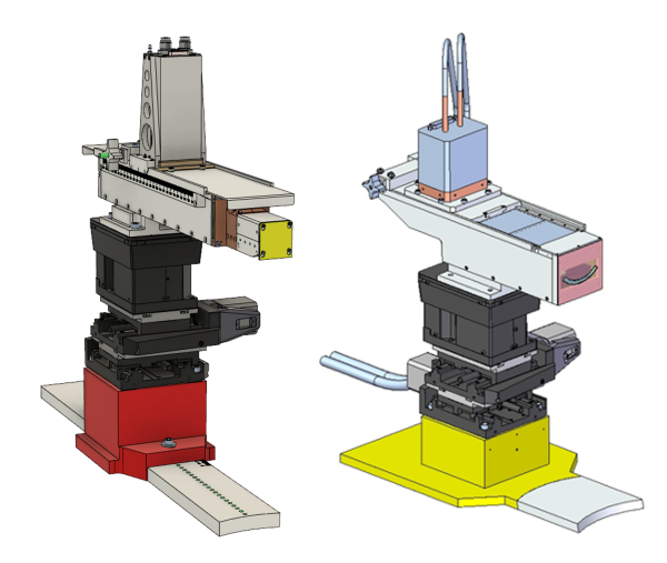
MEC provides standard spectrometers for use in X-ray Thomson Scattering from dense plasmas or compressed targets. These spectrometers use the von Hamos geometry: a cylindrically curved crystal produces a line focus with the measured X-ray spectrum dispersed along the line, and captured onto a CCD. Possible crystal choices include highly oriented pyrolytic graphite (HOPG), germanium, and silicon. Starting from run 18, the LCLS X-ray polarization is vertical, and the spectrometers are designed to operate in the horizontal plane; changes to the observed k-vector must be done manually. MEC has available two standard spectrometers covering the photon range from 4-8 keV, and one covering the range of 8-24 keV.
MEC X-ray Imager
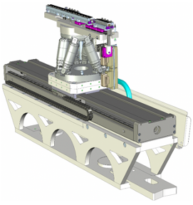
This in-house system can be used to image phenomena with spatial resolution better than 500 nanometers and temporal resolution better than 100 femtoseconds. It was designed for studies relevant to High Energy Density Science, such as imaging shock fronts, phase transitions, void collapses, etc. The in-chamber motorized Be CRL mount known as the MEC X-ray Imager (MXI) flexibly positions up to 3 lens stacks over a wide range within the chamber. It is combined with a flight tube with an imaging platform at the end that uses a YAG screen and various optical microscopes, including an Optique Peter, or else direct detection on various X-ray cameras. The standard imaging plane position for the optics platform is ~4.2 m past the target. The MXI capabilities are published in Scientific Reports (link to article).
X-ray Transmission Crystal Spectrometer (XTCS)
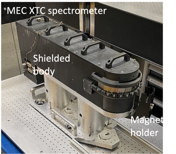
This spectrometer has been designed to enable high resolution hard X-ray spectroscopy in harsh EMP and bremsstrahlung environment, as those found during the interaction of the high intensity laser with solid matter. It uses a Cauchois quartz crystal (the crystal plan are perpendicular to the crystal surface) cylindrically bent allowing the insertion of tungsten slits at the focus of the rays. The spectrometer usually covers >1keV spectral range centered on the central photon energy ranging from 6 to 21 keV at high resolution. When low resolution is needed, the spectral range and throughput is greatly increased at the expense of the spectral resolution. The weight of the fully shielded device is about 350 lbs but can be slid on the optical table and is compatible with all the standard platforms.
Target Chamber
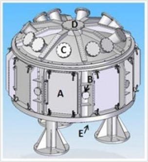
The MEC target chamber is a cylindrical vacuum vessel of 1 inch aluminum that operates in the high vacuum regime using turbo pumps. It has 10 top port, 8 side port, and 6 doors, all oriented towards target chamber. It contains an aluminum breadboard of approximately 2 m diameter, with a 1 inch ¼-20 bolt pattern. The breadboard legs extend to the feet of the vessel and are otherwise isolated from the chamber walls, minimizing shifting during pumpdown. In the middle of the chamber there is a motorized target alignment stage with 6 degrees of freedom.
FDI
Note: the FDI diagnostic is decommissioned. Please contact an instrument scientist if you are interested in having it available for a future run.
The primary object of the Fourier Domain Interferometer (FDI) diagnostic is to measure the phase and amplitude of the reflection of a femtosecond laser of the target. The phase information is extracted, by interfering two time-delayed pulses (one typically before, and the other after an incident pump laser pulse) in a spectrograph and gives information about the motion of the critical density surface of the target. The FDI at MEC is based on a design that originates from LULI, Ecole Polytechique, Paris. It has a time resolution of 35 fs, and a spatial resolution of 10 µm, and can be used in a chirped configuration.
XUV Spectrometer

Note: The XUV spectrometer is decomissioned. Please contact an MEC instrument scientist if you are interested in having it available for a future run.
The XUV Spectrometer for the Matter in Extreme Conditions (MECI) instrument is a diagnostic instrument for MECI experiment to resolve emissions in the XUV regime. It sits inside the MEC target chamber, has a high collection efficiency, wavelength range of 7-35 nm, and resolution of 0.08 nm. It is based on a design by DESY and the University of Jena (R.R. Fauestlin et al, J. Inst., 5, p02004).
LUSI Pulse Picker
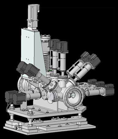
A single pulse shutter is used to allow only a single FEL pulse to pass through to the experimental chambers. A millisecond shutter from azsol GmbH is incorporated into a vacuum chamber on a translation stage to allow insertion into the beam. It can be operated up to 10 Hz.
LUSI Attenuators
A set of silicon foils of varying thicknesses is used to tailor the intensity of the LCLS beam. Multiple attenuation factors is possible by introducing any desired combination of foils into the LCLS beam path. Ten foils of different thicknesses is provided and can be used in any combination.
LUSI Guard Slits
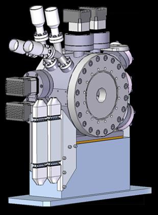
Cylinders of 3 mm diameter made of silicon nitride (Si3N4) and or Tungsten is used to slit the beam. Silicon nitride will not get damaged by the LCLS beam downstream of the Near Experimental Hall, while the Tungsten slits behind the Silicon nitride will remove the Higher harmonics.
LUSI Pop-in Profile-Intensity Monitors
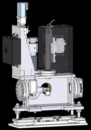
The spatial profile of the LCLS beam is measured at various locations along the MEC beamline using a scintillating screen and a high resolution camera-lens combination. The screen is mounted on a translation stage to allow insertion into the beam. The beam profile measurement is destructive of the beam and is used for alignment and troubleshooting procedures.
LUSI Pop-in Intensity Monitor
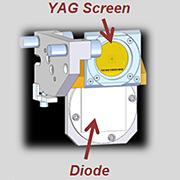
The integrated intensity of the LCLS beam is measured at various locations along the MEC beamline using a photodiode which is mounted on a translation stage to allow insertion into the beam. The intensity measurement is destructive of the beam and is used for alignment and troubleshooting procedures.
LUSI Intensity-Position Monitor
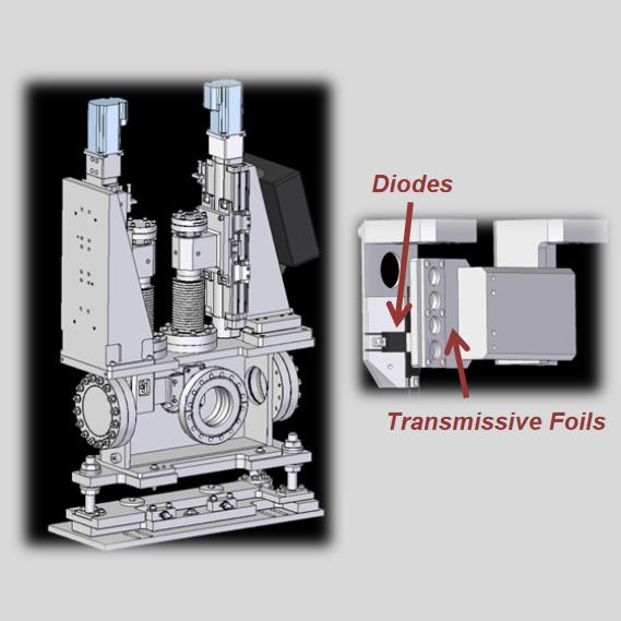
A thin foil allowing most of the beam to be transmitted is used to measure the LCLS pulse energy on a shot-to-shot basis. Compton back-scattering from the thin foil is measured using a set of diodes located upstream of the foil. The sensing area of the diodes is facing the foil and they is place in a tiled arrangement leaving a hole in the middle. The integrated intensity of all the diodes provide a measurement of the beam intensity on every pulse. The relative signal from each tile is used to get a measurement of the beam position.
LUSI X-ray Focusing Lens system
Compound refractive lenses made of Beryllium is used to produce a 1 µm focus in the MEC Target chamber. An translation stage allows one of three stacks of lenses to be selected which allows focusing of photon energies from 4 to 8 keV. The lenses is approximately 4 m from the target chamber center. Focusing below 4 keV is in principle possible but incurs a large intensity penalty due to the absorption below this energy.

|

|
Time Tool
Synchronization of the short pulse laser is performed by spatial encoding of the arrival time of the X-rays backilluminated by the compressed optical laser. The collimated X-rays impinge on a thin piece of sillicon nitride membrane exciting electrons into the valence band. This non-destructive excitation temporarily change the index of refraction of the material to the short pulse optical laser wavelenght which thus becomes opaque when traversing the material. As the silicon nitride foil is placed at 45° to the incomping X-rays, the X-rays first change the index of refraction at the top of the foil, while the bottom sees a change only later, when the bottom part of the X-ray pulse arrives on the target. This geomtry spatially encodes the arrival time and allows non-intrusive sub-ps shot-to-shot measurement of the jitter or drift in the pulses syncrhonization.
MEC CONTACTs
Eric Galtier
MEC Instrument Lead
(650) 926-6227
egaltier@slac.stanford.edu
Ariel Arnott
Area Manager
(650) 926-2604
amarnott@slac.stanford.edu
Gilliss Dyer
MEC Department Head
(650) 926-3414
gilliss@slac.stanford.edu
Hae Ja Lee
Lead Scientist
(650) 926-2049
haelee@slac.stanford.edu
Philip Heimann
Senior Scientist
(650) 926-8772
paheim@slac.stanford.edu
Dimitri Khaghani
Staff Scientist
(650) 926-5009
khaghani@slac.stanford.edu
Bob Nagler
Staff Scientist
(650) 926-3810
bnagler@slac.stanford.edu
Nick Czapla
Associate Laser Scientist
(650) 926-4314
nczapla@slac.stanford.edu
Nina Boiadjieva
Staff Engineer
(650) 926-4035
ninab@slac.stanford.edu
Peregrine McGehee
Staff Engineer
(650) 926-1631
peregrin@slac.stanford.edu
Philip Hart
Staff Engineer
(650) 926-2813
philiph@slac.stanford.edu
Marc Welch
Staff Engineer
(650) 926-3754
mwelch@slac.stanford.edu
Jonathan Ehni
Science & Engineering Associate
(650) 926-4562
jonehni@slac.stanford.edu
-
MEC Control Room
(650) 926-7970
MEC Hutch
(650) 926-7974
Vestibule
(650) 926-7976
MEC LOCATION
